
What is Neuroimaging?
Neuroimaging is a revolutionary field where advanced computational powers of cutting-edge technology are mixed with neuroscience to get high-resolution images of the central nervous system or the human brain. These high-resolution images are later on used to study different parts of the brain and also to locate defects and illnesses present.
Its increasing role is also responsible for increasing numbers of researches and hence unveiling the truths, myths and complexities of brain. As neuroscience is a multidisciplinary field comprising knowledge of computer science, biological chemistry, neuroscience, psychology, and statistics, research in this field is not a child’s play. As a result of this research and update in this field, new complexities have been revealed, and neuroimaging has become an invaluable tool for researchers, clinicians, and scientists seeking to decode the mysteries of cognition, behavior, and neurological disorders.

Table of Contents
Neuroimaging Techniques
Brain imaging techniques that are used are classified as:
- Structural Imaging
- Functional Imaging
- Diffusion Imaging
- Molecular Imaging
1.Structural Imaging
Structural neuroimaging is a category of imaging techniques that is specialized for visualization of anatomical and morphological images of brain. These techniques provides detailed image of brain’s physical form, as a result allowing researchers and clinicians to examine its various regions, and to identify abnormalities present. Structural neuroimaging is valuable for understanding the brain’s architecture and diagnosing conditions that affect its integrity.
The primary objective of structural neuroimaging is to create high-resolution images of different structure of brain that reveals size, shape and composition of its structures. The most common structural neuroimaging techniques are:
- Magnetic Resonance Imaging(MRI)
- Computed Axial Tomography(CT)
- Cranial Ultrasound Imaging
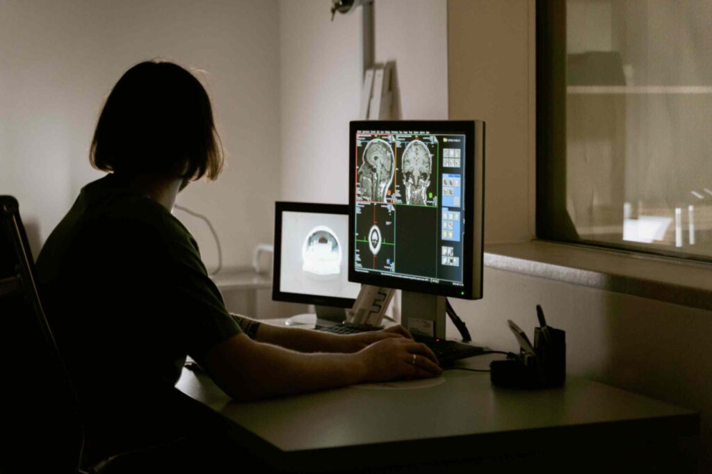
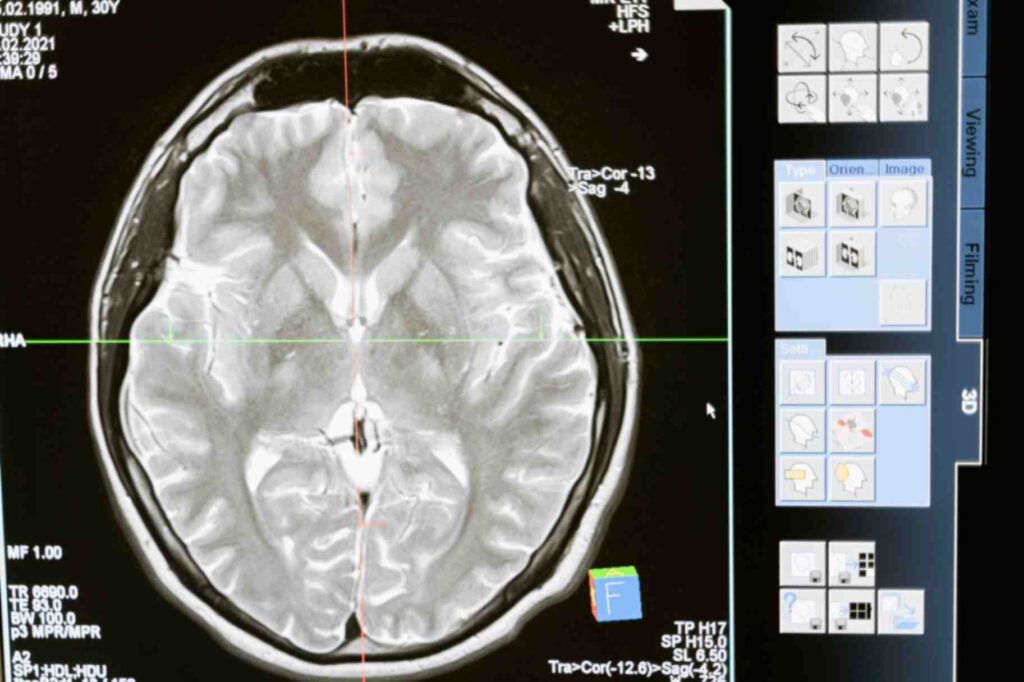
1.Magnetic Resonance Imaging(MRI)
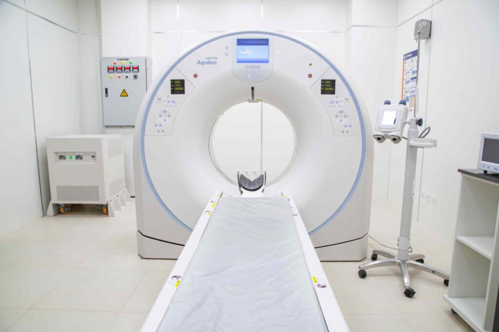
MRI is a non-invasive imaging technique that uses strong magnetic fields and radio waves to generate detailed two or three dimensional images of brain’s internal structures without ionizing them. This technique produces images with high spatial resolution, allowing visualization of different regions of brain, such as the cortex, subcortex and cerebellum.
Images produced by MRI technique are used in setting of diagnostic plans for conditions like tumors, developmental disorders, and vascular abnormalities.
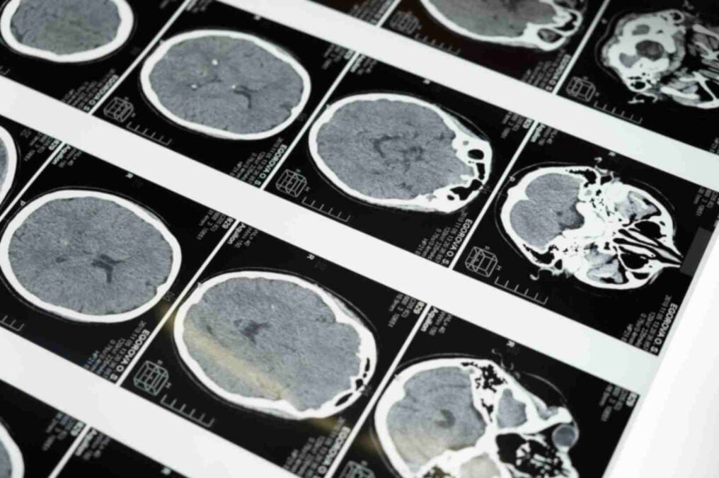
2.Computed Axial Tomography(CT)
CT scans uses X-rays from different directions which creates detailed images of the brain. This method of imaging is not frequently used as MRI for structural neuroimaging. CT scan is based on a computer software that performs a numerical integral calculation on the number of X-rays beam absorbed that provides the amount of X-rays absorbed in a small volume of brain and so it is quick method of neuroimaging as compared to MRI and hence CT scans become valuable in emergency situations to quickly assess traumatic brain injuries like skull fractures. CT scan provides a rapid overview of the brain’s structure, making it more useful in acute medical settings.
3.Cranial Ultrasound Imaging
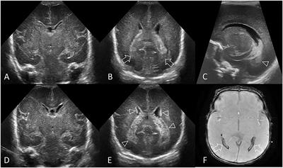
Image Credit : Frontiers
In certain cases, ultrasound imaging is employed to visualize the brain which is usually only only used in babies, as their fontanelles provides gap for ultrasound that makes ultrasound imaging possible for infants. This method of imaging is highly portable but the images produced through this technique produces lower resolution as compared to MRI.
2.Functional Imaging
Functional neuroimaging refers to a set of techniques used to image brain and observe brain activity while individuals engage in cognitive tasks or experience stimuli. These techniques provides insights of neural mechanisms underlying various cognitive functions, emotions, behaviors. Functional neuroimaging methods are crucial in mapping brain regions associated with specific tasks, and investigating disorders or abnormalities. The most common functional neuroimaging techniques include:
- Functional Magnetic Resonance Imaging (fMRI)
- Positron Emission Tomography (PET)
- Electroencephalography (EEG)
- Magnetoencephalography (MEG)
- Near-Infrared Spectroscopy (NIRS)
1.Functional Magnetic Resonance Imaging (fMRI)
One of the widely used functional neuroimaging techniques that measures blood flow changes in brain, which are associated with neural activity. This method images with high spatial resolution of around 2-3 millimeters and the method is non-invasive. The principle of this test is based upon paramagnetic properties of oxygenated and de-oxygenated hemoglobin that shows pattern of blood flow in the brain which is used for imaging purpose.
2.Positron Emission Tomography (PET)
PET imaging technique relies on a foreign radioactive tracer which needs to be injected in a small amount in the bloodstream. The tracer used have very short half-life span of about ~2 hours, and as it decays, it emits positrons, and the resulting gamma rays are detected.
This technique measures regional cerebral blood flow, metabolism, or neurotransmitter receptor binding. Currently fMRI method of imaging is preferred over PET scans as it does not involves injection of foreign tracer or radiation, and has a high temporal resolution as compared to PET scans.
3.Electroencephalography (EEG)
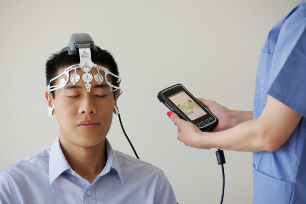
One of the neuroimaging technique that provides excellent temporal resolution, in this technique a set of electrodes are placed over the scalp that provides Event-related potentials (ERPs) on response to a specific stimuli or event. With the help of these ERPs researchers and clinicians can examine the exact timing of different neural events that can be used in clinical applications.
4.Magnetoencephalography (MEG)
MEG measures the magnetic field produced by electrical activity of neurons. Extremely sensitive devices like SERF and SQUIDs magnetometers are used to measure these neuronal activity, although images produced by this method has high temporal resolution but spatial resolution is low as compared to EEG technique.
The main objective of using this technique is magnetic fields tend to get less by surrounding tissues of the head, but this is not the case with electrical signals used in EEG technique. Hence as an outcome of this MEG technique becomes an invaluable resource for researchers and clinicians in molding the skull in about a collection of spherical shells.
5.Near-Infrared Spectroscopy (NIRS)
NIRS method of imaging uses near-infrared light that is passed through the scalp, measures the change blood oxygen level in brain through detecting reflected light.
This method is very portable with easy setting methods that makes it suitable for certain clinical applications.
3. Diffusion Imaging
Diffusion imaging is a technique which is used to study diffusion of water molecules in biological tissues, especially is brain. This imaging technique provides information about microstructures and organization of white matter tracts. This helps to investigate the neural connection and the neural pathways.
This method focuses primarily on detecting and hence allowing researchers to study about the movement of water molecule in brain which is influenced by some structural features of brain tissues
Some of the most common terms in diffusion neuroimaging techniques are
Mean Diffusivity(MD): It is a metric used to represent average diffusion of water molecules without taking direction into consideration.
Fractional Anisotropy(FA): It is a value used to represent degree of anisotropy and directionality of water diffusion in tissues. A high value of FA represents more directional and organized diffusion in tissues.
The most common diffusion imaging techniques include
- Diffusion Tensor Imaging (DTI)
- Diffusion Kurtosis Imaging (DKI)
- High Angular Resolution Diffusion Imaging (HARDI)
1.Diffusion Tensor Imaging (DTI)
DTI is a special type of neuroimaging technique assigned to measure three-dimensional diffusion of water molecules in tissues. This method is particularly useful in mapping white matter tracts in the brain often used in studying brain connectivity.
2.Diffusion Kurtosis Imaging (DKI)
An extension method of DTI employed to provide information about tissue microstructure along with the mapping of white matter tract.
3.High Angular Resolution Diffusion Imaging (HARDI)
HARDI is a more advanced and complex diffusion imaging technique that uses a large number of direction of diffusion to capture more complex orientation of fibers. It is particularly useful in region where fiber branching or crossing occurs.
Some of the major applications of diffusion neuroimaging technique includes
- Brain connectivity disorders
- White Matter Tractography
- Neurological and Psychiatric Disorders
4.Molecular Imaging
Molecular neuroimaging technique is a specialized field within neuroimaging that focuses on visualizing and studying molecular processes in brain. This technique assess molecular and biomolecular functions, providing insights about neurotransmitter system, metabolic processes and receptor binding.
Molecular neuroimaging is particularly more usable in the study of neurodegenerative disorders like Alzheimer’s disease, Parkinson’s disease and psychiatric disorders like depression and schizophrenia, and in the development and evaluation of new pharmacological interventions.
Its application has a crucial role in both clinical and research fields. Its major applications include
- Metabolic Imaging
- Pharmacological Studies
- Receptor Binding Studies
- studying neurotransmitter Function
The most common methods of molecular neuroimaging are
- Positron Emission Tomography (PET)
- Single-Photon Emission Computed Tomography (SPECT)
- Magnetic Resonance Spectroscopy (MRS)
1.Positron Emission Tomography (PET)
It is a powerful neuroimaging technique that uses radiotracers labeled with positron-emitting isotopes. These radiotracers are injected into the body, and their distribution is detected by PET scanners that is used to study numerous of molecular processes.
2.Single-Photon Emission Computed Tomography (SPECT)
SPECT is another nuclear imaging technique that uses gamma-emitting radiotracers. As like the PET technique this technique also allows the imaging of molecular processes.
3.Magnetic Resonance Spectroscopy (MRS)
This is a non-invasive molecular imaging technique that uses MRI to detect and quantify specific type of chemical compounds in the brain. It provides information about neuronal health and metabolism.
FAQs on neuroimaging
How to get into neuroimaging?
Start by earning a bachelor’s degree in a relevant field such as neuroscience, psychology, biology, physics, engineering, or a related discipline. Make sure to include coursework in mathematics and computer science. Work in research labs or participate in internships to gain hands-on experience in neuroscience or imaging-related research. Consider pursuing a graduate degree (master’s or Ph.D.) in a field related to neuroimaging. Programs in neuroscience, biomedical engineering, medical physics, or a related discipline can provide the necessary background. Take courses specifically focused on neuroimaging techniques, such as MRI, fMRI, PET, SPECT, or diffusion imaging. Familiarize yourself with data analysis methods and tools used in the field.
Look for internships or fellowships that provide practical experience in neuroimaging. Many research institutions and imaging centers offer opportunities for hands-on training.
How to become neuroimaging technician?
Obtain an associate degree or certification in radiologic technology or a related field. Complete on-the-job training in neuroimaging techniques and equipment. Obtain relevant certifications, gain experience through internships or entry-level positions.
How much neuroimaging costs ?
Cost of neuroimaging varies from place to place and country to country, it also depends on factors like type of imaging procedure you are going for, type and facilities of hospitals you are going for etc.
How to do functional neuroimaging?
A detailed explanation of functional neuroimaging, including its techniques are given in the article, kindly refer above.
How accurate is neuroimaging?
Accuracy of neuroimaging depending on the type of technique you are going for. But it can be said that it is quite accurate that it is able to give relevant information about your brain to the clinicians.
What is neuroimaging used for?
Certain use-cases of neuroimaging are
- Research in Neuroscience
- Clinical Diagnosis and Assessment
- Mapping Brain Connectivity
- Treatment Planning and Monitoring
- Pharmacological Studies
- Cognitive and Behavioral Research
- Traumatic Brain Injury (TBI) Assessment
Is X-ray a neuroimaging technique or not?
No, X-ray technique is not considered as a neuroimaging technique, as it is primarily applicable to visualization of bony structure of the skull and also it does not give information about soft tissues of brain.
Is EEG a neuroimaging technique or not?
Yes, EEG is a neuroimaging technique. You can read more about EEG this article above.
Structural vs functional neuroimaging.
A detailed explanation of of structural and functional neuroimaging techniques is given kindly refer.
what is pediatric neuroimaging?
Pediatric neuroimaging is a specialized field of medical imaging focused on the diagnosis and assessment of neurological conditions in children. It involves the use of various imaging techniques to visualize and study the structure and function of the developing brain and spinal cord in pediatric patients. The goal is to identify and understand abnormalities, developmental issues, and diseases affecting the pediatric nervous system.
Read my article on laptops under 60k
2 thoughts on “Neuroimaging:14 Brain imaging methods”